
MASD: What Are the Types of Moisture-Associated Skin Damage?
Practice Accelerator
February 1, 2018
February 1, 2018
Keywords
Categories
It has long been known in clinical practice that long-term exposure of the skin to moisture is harmful and can lead to extensive skin breakdown. The term moisture-associated skin damage was coined as an umbrella term to describe the spectrum of skin damage that can occur over time and under various circumstances. To have a moisture-associated skin condition, there must be moisture that comes in contact with that skin. Moisture-associated skin damage or MASD is defined as "inflammation and erosion of the skin caused by prolonged exposure to moisture and its contents, including urine, stool, perspiration, wound exudate, mucus, or saliva."1,2 MASD encompasses the four forms of moisture-associated skin damage, which include 1) incontinence-associated dermatitis or IAD; 2) intertriginous dermatitis, also called intertrigo or ITD; 3) peristomal moisture-associated dermatitis; and 4) periwound moisture-associated dermatitis. Now let's take a closer look at each one.
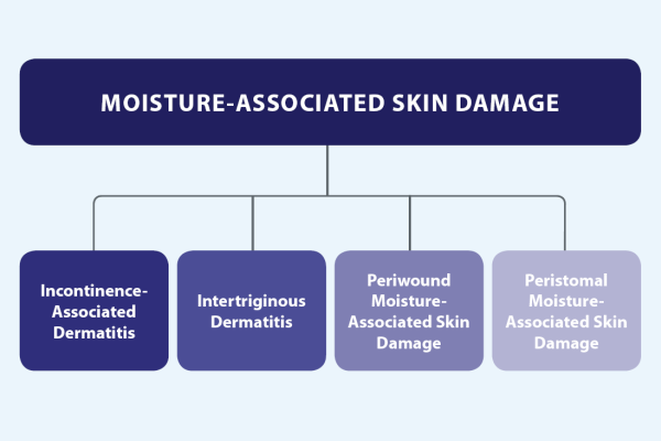
The types of moisture-associated skin damage
Incontinence-Associated Dermatitis
The first type of MASD is incontinence-associated dermatitis or IAD, which has many aliases such as diaper dermatitis, maceration, diaper rash, perineal dermatitis, and moisture lesions. IAD develops as a form of irritant dermatitis resulting from prolonged or chronic exposure to urine and/or stool, particularly liquid stool. These lesions are also often mistakenly labeled stage 1 or stage 2 pressure ulcers/injuries.3 IAD occurs in the perineum, including the labial folds and vulva to the anus in women, and from the scrotum to the anus in men; groin, buttocks, gluteal cleft, and even extending down to the inner and posterior thighs.4 When the skin is moist, it is more susceptible to damage from pathogens. Skin damage may be exacerbated by the use of alkaline cleansers, soaps or detergents, aggressive cleaning, and inappropriate use of containment devices.
Intertriginous Dermatitis
Now let's look at intertriginous dermatitis, also called intertrigo or ITD, which is an "inflammatory dermatitis of opposing skin surfaces caused by moisture accumulation in skin folds and friction between the opposing skin surfaces."4 This can occur more often among the bariatric population, those with diabetes mellitus, and those on bed rest. ITD can also be associated with containment devices or any other types of medical devices where moisture may be trapped in a skin fold. Some of the most common locations for ITD are the groin, the axilla, underneath the breast, and in the abdominal panniculus or pannus. It is important to consider every skin fold as having the potential for skin breakdown. ITD can manifest with erythema, denudation, itching, oozing, or weeping. An alteration in the normal skin flora may also contribute to the overgrowth of Candida, for example, or for another fungal infection or a bacterial overgrowth.5 The importance of checking all skin folds cannot be overemphasized.
Peristomal Moisture-Associated Dermatitis
Another form or MASD is peristomal moisture-associated dermatitis, which is an inflammation or erosion of the skin related to moisture from fecal, urinary, or chemical irritants beginning at the stoma skin junction, which can extend outward in approximately a four-inch radius.6 The location of the stoma and the type and consistency of the effluent may have an impact on the potential for breakdown of the peristomal skin and may also affect the treatment choices for protecting and treating the skin. Despite the utilization of various containment devices and strategies, in individuals with hyperactive bowels, diarrhea, or fistulas that connect the bowel and the skin, fecal effluent may leak out onto and erode peristomal skin. Obtaining and maintaining a secure fit with a pouching system are the biggest challenges.
Poor appliance fit and adherence contribute to leakage and erosion of the epidermis, which in turn perpetuate the vicious cycle of inability to maintain secure pouching post maceration. The issue of peristomal moisture-associated dermatitis also exists for other ostomy sites. For instance percutaneous gastrostomy tube insertion sites have the potential for leakage of digestive enzymes and tube feeding around the tubing. Patients with tracheostomies may develop skin issues resulting from perspiration, saliva, or sputum accumulation around the stoma, under the flange of the external cannula, or on the tracheostomy ties or dressing.7
Periwound Moisture-Associated Dermatitis
The fourth type of MASD is periwound moisture-associated dermatitis, and it occurs when wound exudate, which contains proteolytic enzymes, comes in contact with periwound skin for prolonged periods of time. The exudate of chronic wounds can exacerbate the problem because it contains a higher concentration of bacteria and its released histamines, as well as proteolytic enzymes known as matrix metalloproteases or MMPs. The lower the viscosity of wound exudate, the lower the protein content will be, hence a lower amount of proteolytic enzymes. 8 Prolonged exposure of periwound skin to the excessive moisture plus these enzymes leads not only to maceration, but also to excoriation, which Dowsett, Groemann, and Harding defined as an injury to the skin caused by trauma, chemical, or thermal burn.9 This excoriation can stem from not just the impact of the enzymes, but also from the physical scratching of the area by the patient because of associated irritation and itching. The skin is then more susceptible to pressure, shear, and friction as well, which can prevent the wound from closing.9
Prevention and Treatment of Moisture-Associated Skin Damage
Prevention and treatment of MASD begin with a thorough assessment of the skin. Proper, yet gentle cleansing of the skin should occur as soon as possible after soiling. Cleansers should be pH balanced and contain gentle surfactants that protect the lipid profile of the skin. No-rinse formulas will allow for cleansing without requiring additional friction. Once the skin is cleansed, barrier products should be applied to repel/protect the skin from subsequent episodes of prolonged exposure to moisture and other caustic agents. Taking MASD into account, emollients may be used to aid in retaining moisture in the stratum corneum by rehydrating the corneocytes.
This will also help prevent transepidermal water loss or TEWL.1 Remembering the fundamental rules of cleanse, moisturize, and protect will not only aid in the repair of damaged skin but also lay the foundation for keeping skin intact and preventing breakdown.
References
1. Young T. Back to basics: understanding moisture-associated skin damage. Wounds UK. 2017;13(4):56-65.
2. Gray M, Black JM, Baharestani MM, et al. Moisture-associated skin damage: overview and pathophysiology. J Wound Ostomy Continence Nurs. 2011;38(3):233-41.
3. Kottner J, Blume-Peytavi U, Lohrmann C, Halfens R. Associations between individual characteristics and incontinence-associated dermatitis: a secondary data analysis of a multi-centre prevalence study. Int J Nurs Stud. 2014;51(10):1373-80. doi.org/10.1016/j.ijnurstu.2014.02.012.
4. Black JM, Gray M, Bliss D, et al. MASD part 2: incontinence-associated dermatitis and intertriginous dermatitis. J Wound Ostomy Continence Nurs. 2011;38(4):359-70. doi:10.1097/WON.0b013e31822272d9
5. Bryant RA. Types of damage and differential diagnosis. In: Bryant RA, Nix DP, eds. Acute and Chronic Wounds: Current Management Concepts. 5th ed. St. Louis, MO: Elsevier; 2016:82-108.
6. Gray M, Colwell JC, Doughty D, et al. Peristomal moisture-associated skin damage in adults with fecal ostomies: a comprehensive review. J Wound Ostomy Continence Nurs. 2013;40(4):389-99.
7. Woo KY, Beeckman D, Chakravarthy D. Management of moisture-associated skin damage: a scoping review. Adv Skin Wound Care. 2017;30(11):494-501. doi: 10.1097/01.ASW.0000525627.54569.da.
8. Voegeli D. Incontinence-associated dermatitis: new insights into an old problem. Br J Nurs. 2016;25(5):256-62.
9. Dowsett C, Groemann NM, Harding K. Taking wound assessment beyond the edge. Wounds Int. 2015;6(1):19-23.
The views and opinions expressed in this content are solely those of the contributor, and do not represent the views of WoundSource, HMP Global, its affiliates, or subsidiary companies.
More from this Author
// fixed missing link variable.
// fixed missing link variable.
// fixed missing link variable.


