Wound Bed Preparation Challenges
June 30, 2021
Introduction
Wound bed preparation has been performed for decades in managing wounds of various etiologies. The wound healing process consists of a complex interlinked and independent cascade, which not all wounds follow in a consistent, organized manner. The TIMERS acronym, consisting of four general steps, has assisted clinicians globally to provide a systematic approach to wound bed preparation that includes Tissue debridement, Infection or Inflammation, Moisture balance, Edge effect, Regeneration and repair, and Social factors.1 Clinicians should have practical knowledge of the principles of advanced wound care management, as well as the challenges faced in treating complex wounds.
Patient Factors and Contraindications
Wound bed preparation is important for wound healing, especially in hard-to-heal wounds. These wounds fail to progress through the normal healing process. Including a systematic approach to wound bed preparation provides the necessary framework for a structured approach to wound healing and leads to better healing outcomes.1 Sometimes certain steps of wound bed preparation are difficult to implement because of patient factors such as diabetes, anemia, malnutrition, immunodeficiency, age, obesity, and drug use or smoking. Contraindications also create challenges in wound bed preparation, including wound etiology, medications, and pain.
How much do you know about wound bed preparation? Take our 10-question quiz to find out! Click here.
Debridement Methods
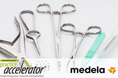
Debridement is necessary in chronic wounds that contain devitalized tissue or biofilm, for preventing infection, and in promoting re-epithelialization.2,3 There are various methods of debridement that can be used alone or in conjunction to optimize the healing environment. These methods include biological, enzymatic, autolytic, mechanical, surgical or sharp, hydrosurgical, synergistic, and ultrasonic.3 Beginning appropriate debridement early is likely to accelerate healing and reduce wound management costs.4 It is important that clinicians are aware of situations when sharp debridement is contraindicated, more difficult, or even impossible. Licensed health care professionals should have practical knowledge and skills to identify the appropriateness of most debridement methods. Selecting one or more debridement methods helps enhance healing, prevents infection, and improves patients’ outcomes. Importantly, licensed health care professionals should know their own scope of practice, requisite skills, and when to refer to patients the most appropriate specialists.
Wound Etiology
A wound’s etiology can have an impact on the types of treatments used. Knowing what caused the wound can help clinicians to select treatments that will help the wound to heal, rather than doing more damage. Pyoderma gangrenosum (PG)–type wounds with devitalized tissue should be treated with an enzymatic debriding agent. These wounds can quickly progress to a large ulceration and be very painful.5 Sharp debridement is contraindicated for PG because it can cause the wound to worsen. Calciphylaxis wounds with expanding tissue necrosis and violaceous borders should not be debrided. Patients must complete sodium thiosulfate therapy and undergo clinical observation to ensure that necrosis expansion and a violaceous border are no longer present before beginning debridement.6 Autoimmune wound types tend to worsen with sharp debridement when there is a prominent active border.
Wound Location
Dry, stable, intact eschar in ischemic limbs (heels) should not be removed because of blood flow in the underlying tissue and high susceptibility to infection.7
Medications
Anticoagulant medications should be closely monitored, and the physician may have the patient withhold anticoagulant therapy based on the nature of the procedure (conservative sharp debridement, surgical sharp debridement).8
Pain Tolerance
Pain assessment and monitoring should be a part of wound management. Not all patients have the same pain tolerance. Communicate with patients about their pain intensity, location of pain, pain patterns, pain triggers, self-treatments, side effects (physical, emotional, psychological), and whether or not they are experiencing pain during treatment. Consider the patient’s needs in creating an individualized pain management regimen.9
Palliative and Terminally Ill Patients
In patients receiving palliative care or patients who are terminally ill, quality of life should come first when deciding on whether sharp debridement is appropriate.10 Other methods of debridement, such as autolytic and enzymatic, are less aggressive and may be more appropriate in these populations.
Infection Management
All wounds are at risk for localized or systemic infection. Wound cleansing is an essential part of wound bed preparation and will help reduce bacteria and foreign debris. Monitor wounds routinely for suspicion of localized biofilm and classic signs and symptoms of infections. All chronic wounds, regardless of etiology, have bacteria, and most chronic wounds contain bacteria and fungi living within a bioconstruct.11 Combining debridement methods has been found to provide an advantage in managing complex wounds with pathological issues.12
The most important step in chronic wound care is to remove necrotic tissue and the microbial bioburden by surgical or sharp debridement.13 Practical knowledge of biofilm prevention and reformation and identification of bacterial balances are essential in optimizing wound healing outcomes. Wounds in proximity to the products of incontinence should be managed and monitored closely because of the significant impact on skin integrity. This can be a challenge for the patient and clinician, but it is a vital step in preventing further breakdown and infection. In patients with both a wound and incontinence, there should be an incontinence management plan in place, including incontinence assessment, routine skin inspection, an effective skin cleansing regimen, skin protection, treatment of incontinence, and education for the patient.14
Moisture Balance
Moisture control is vital in wound healing of all wound types. An optimal moist wound healing environment facilitates the action of growth factors, cytokines, and chemokines, thereby enhancing the cellular growth needed in setting the stage for tissue repair. When the skin is exposed to prolonged moisture, it becomes macerated and more susceptible to skin impairment and damage. Urine and feces change the skin environment and allow bacterial and fungal growth. When maintaining skin integrity, it is critical to keep the skin clean and dry; this may include barrier creams, gentle cleansers, absorbent dressings, moisturizers, incontinence products, and fecal management systems.15
Desiccation
Wounds that are desiccated (extreme dryness) also experience delayed healing. The wound bed cells dehydrate and offset the healing process. Wounds that maintain a moist wound healing environment stimulate cell migration and move toward wound closure. Hydrogel-based wound care dressings donate moisture and are commonly used for hydrating wounds.
Conclusion
Wound bed preparation practices will help create an optimal wound healing environment leading hard-to-heal wounds toward a healing trajectory. Wound bed preparation is vital for wound healing, but it is not always a simple process. Always evaluate the wound to see what areas need addressing—moisture, devitalized tissue, rolled edges—to create a plan of care that resolves all stalling factors.

References
- Halim AS, Khoo TL, Saad AZ. Wound bed preparation from a clinical perspective. Indian J Plast Surg. 2012;45(2):193-202. doi:10.4103/0970-0358.101277
- Frykberg RG, Banks J. Challenges in the treatment of chronic wounds. Adv Wound Care (New Rochelle). 2015;4(9):560-582.
- Manna B, Nahirniak P, Morrison CA. Wound Debridement. Treasure Island, FL: StatPearls Publishing; 2021. https://www.ncbi.nlm.nih.gov/books/NBK507882/. Accessed June 17, 2021.
- Effective debridement in a changing NHS: a UK consensus. Wounds UK. 2013. https://www.wounds-uk.com/resources/details/effective-debridement-in-a-…. Accessed March 25, 2021.
- Pompeo MQ. Pyoderma gangrenosum: recognition and management. Wounds. 2016;28(1):7-13. https://www.woundsresearch.com/article/pyoderma-gangrenosum-recognition…. Accessed March 25, 2021.
- Carpenter S, Shaffett TP. Choosing the best debridement modality to ‘battle’ necrotic tissue: pros & cons. Todays Wound Clin. 2017;11(7).
- Ayello EA, Baranoski S, Sibbald RG, Cuddigan JE. Wound debridement. In” Baranoski S, Ayello EA, eds. Wound Care Essentials: Practice Principles. 4tth ed. Philadelphia, PA: Wolters Kluwer Lippincott Williams & Wilkins; 2012.
- Polania Gutierrez JJ, Rocuts KR. Perioperative Anticoagulation Management. Treasure Island, FL: StatPearls Publishing; 2021. https://www.ncbi.nlm.nih.gov/books/NBK557590/. Accessed June 17, 2021.
- Minimising Pain at Wound Dressing-Related Procedures. A Consensus Document. World Union of Wound Healing Societies' Initiative. London, UK: MEP Ltd; 2004.
- American Medical Directors Association (AMDA). Pressure Ulcers in the Long-Term Care Setting Clinical Practice Guideline. Columbia, MD: AMDA; 2008.
- Attinger C, Wolcott R. Clinically addressing biofilm in chronic wounds. Adv Wound Care (New Rochelle). 2012;1(3):127-132. doi:10.1089/wound.2011.0333
- Kalan L, Loesche M, Hodkinson BP, et al. Redefining the chronic-wound microbiome: fungal communities are prevalent, dynamic, and associated with delayed healing. MBio. 2016;7(5):e01058-16.
- Wolcott RD, Cox SB, Dowd SE. Healing and healing rates of chronic wounds in the age of molecular pathogen diagnostics. J Wound Care. 2010;19(7);272-278, 280-281.
- Cooper P. Skin care: managing the skin of the incontinent patient. Wound Essentials. 2011;6:69-74.
- National Health Service (UK). Great skin. How to: manage incontinence/moisture. https://nhs.stopthepressure.co.uk/How-To-Guides/howtogreatskinincontine…. Accessed March 25, 2021.
The views and opinions expressed in this content are solely those of the contributor, and do not represent the views of WoundSource, HMP Global, its affiliates, or subsidiary companies.


